Macular Hole

The retina is the inside lining of the back of the eye. It is composed of many layers of light sensitive nerve cells. Light entering the eye is focused on the retina and is converted to electrical impulses which are carried to the brain by the optic nerve. The centre of the retina is called the macula. It is the most sensitive part of the retina and is used for day light and colour vision. This is in contrast to the rest of the retina which is more sensitive in the dark and is mainly used for navigational black and white vision at night.

The retina is supplied by one main artery and one main vein which enter through the optic nerve and branch all over the retina to smaller arteries and veins that cross each other like railway tracks.

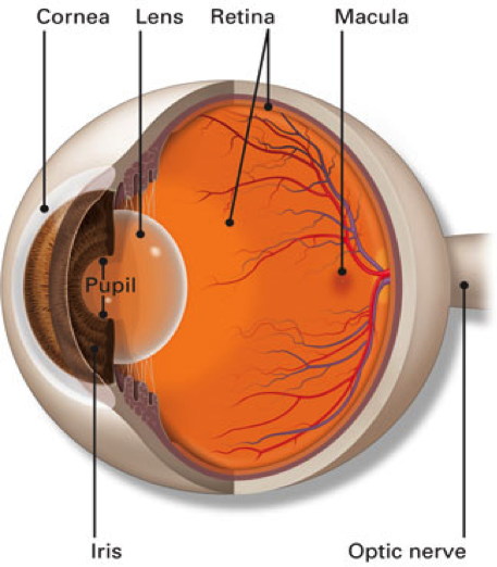
source – American Academy Ophthalmology

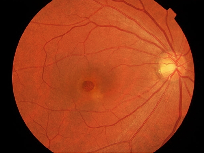
The macula is the thinnest and most sensitive part of the retina located at its centre. We focus an image on the macula to see it clearly and in colour. A macular hole condition occurs when there is a full thickness defect at the macula.

There are stages for macular hole depending on its size and the condition of the vitreous (gel inside the eye). In chronic cases the hole becomes large and more damage is done to the nerve cells of the macula (stage 4)

Retinal Specialist Melbourne

Macular hole-stage 4

  • Spontaneous: in most cases, macular hole occurs without any obvious reason. With normal ageing, the vitreous (gel inside the eye) collapses and pulls on the retina causing the development of a hole at the macula.
  • Trauma: blunt injury to the eye
  • High Myopia (short-sightedness)
  • Macular oedema
  • Epiretinal membrane
  • Sex: females are more likely to develop a macular hole than males
  • Age: macular hole occurs more commonly in people aged 60 and above
  • Difficulty focusing
  • Blind or grey spot in central vision
  • Central distortion of vision
  • Change of colour perception

Peripheral vision always remains intact in the presence of a macular hole. You may be able to focus on an object by looking at it slightly sideways.

  • Dilated retina examination: This is the standard test to check the back of your eye (retina). You will have your pupil dilated with drops so your vision will be blurry for an hour or two after the examination. Our specialist will check your eyes on the microscope (slit lamp) and use a special lens to see the details of the retina. The peripheral retina will be checked with another instrument called an indirect ophthalmoscope. You will also receive a full examination of your eyes including the front of the eye (anterior segment).
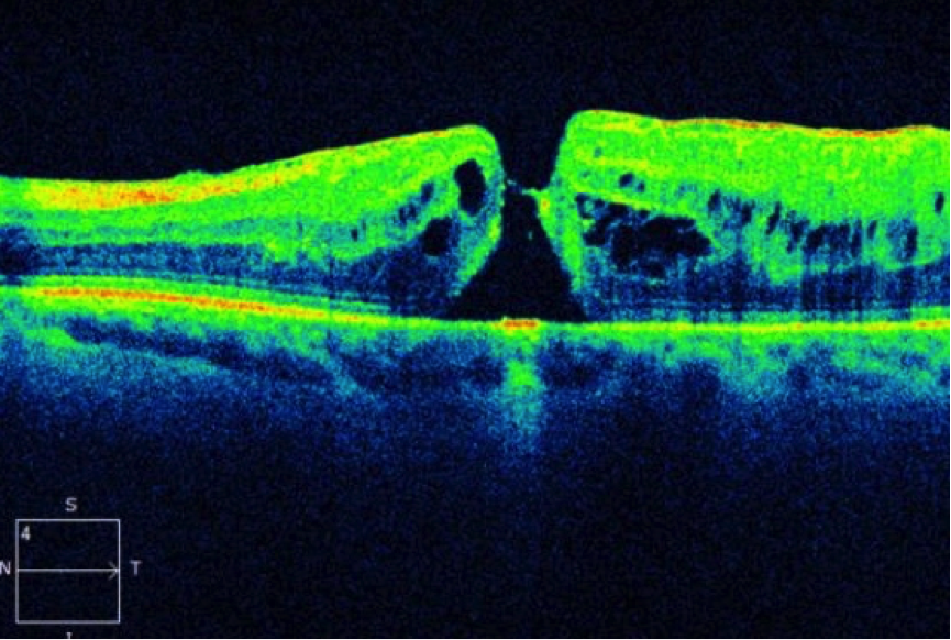
  • OCT (optical coherence tomography): This is a fast scan of the retina centre (macula) which shows cross section details of its layers. This test will clearly identify a break in the retina at the macula.


OCT of macular hole

  • Vitrectomy surgery: Vitrectomy has a 95% success rate in closing macular hole. The surgery is more successful if the condition is diagnosed early and the macular hole is small in size. The procedure includes removal of the vitreous by a special machine using an operating microscope. Afterwards the inner fine lining of the retina (internal limiting membrane) is stained by a special dye and peeled from the macula. At conclusion, the vitreous cavity is filled with a special gas to help close the macular hole. In most cases the patient is asked to keep their face down for a few hours a day over the following 3 to 5 days to insure success of surgery.

Note: if you receive gas inside the eye, you cannot fly or travel to high altitude until the gas has resolved

  • Ocriplasmin (Jetrea): this is a relatively new method of closing the macular hole by a simple injection of a drug into the vitreous cavity. It releases any vitreous traction on the edges of the macular hole to assist its closure. It is used in specific conditions.
  • Observation: This may be indicated for a very small macular hole at early stages as in some cases it can close spontaneously.

Want to schedule an appointment?

Call us at (+61) 3 9459 5133 or fill in the appointment form...