Epiretinal Membrane

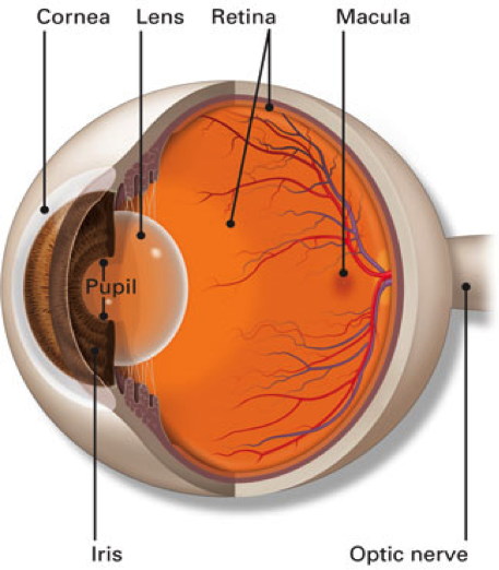
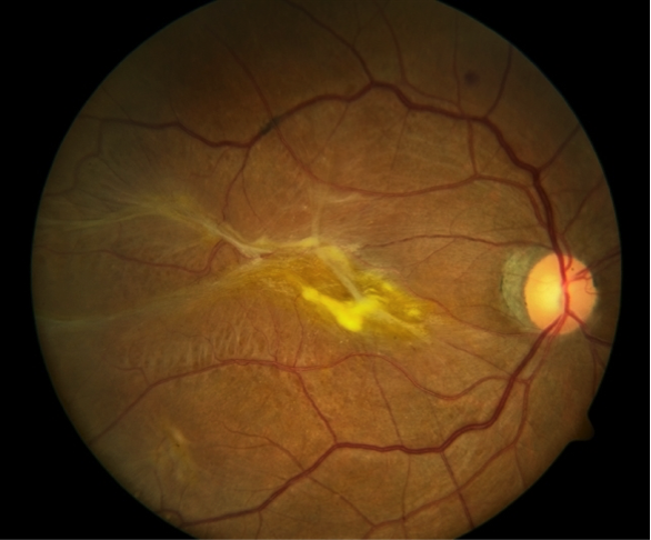
The retina is the inside lining of the back of the eye. It is composed of many layers of light sensitive nerve cells. Light entering the eye is focused on the retina and is converted to electrical impulses which are carried to the brain by the optic nerve. The centre of the retina is called the macula. It is the most sensitive part of the retina and is used for day light and colour vision. This is in contrast to the rest of the retina which is more sensitive in the dark and is mainly used for navigational black and white vision at night.

The retina is supplied by one main artery and one main vein which enter through the optic nerve and branch all over the retina to smaller arteries and veins that cross each other’s like railway tracks.

source – American Academy Ophthalmology

Epiretinal membrane, also called cellophane maculopathy or macular pucker, is a semitransparent tissue that grows on the inner surface of the retina and distorts the retinal surface by dragging it causing it to wrinkle. If it occurs at the macula, centre of the retina, it will affect the vision.

Fundus photo of a dense epiretinal membrane in the right eye
source – American Society of Retina Specialists

  • Idiopathic: No specific cause for the ERM to develop apart from the natural aging process. The vitreous gel changes its structure and separates from the inner retinal surface. This process is called posterior hyaloid detachment. In the space created between the back surface of the vitreous gel and the retina, glial cells start to grow and develop a fine tissue like membrane on the retinal surface.
  • Retinal tears and retinal detachment
  • Eye inflammation
  • Diabetic retinopathy
  • Retinal vein occlusion
  • Trauma
  • Ageing: by the age of 75, 20% of people will have evidence of epiretinal membrane.
  • Posterior hyaloid detachment.
  • No symptoms: many patients will not notice any problems if the epiretinal membrane is mild and only causing mild irregularity of the retinal surface.
  • Distortion (metamorphopsia): if there is considerable wrinkling of the retina surface, straight lines will appear wavy and crooked.
  • Blurred vision; if the membrane grows more dense, central vision will become blurred with loss of definition.
  • Image size changes: images may become smaller (micropsia) or larger (macropsia) than normal.
  • Dilated retina examination: This is the standard test to check the back of your eye (retina). You will have your pupil dilated with drops so your vision will be blurry for an hour or two after the examination. Our specialist will check your eyes on the microscope (slit lamp) and use a special lens to see the details of the retina. Peripheral retina will be checked with another instrument called an indirect ophthalmoscope. You will also receive a full examination of both eyes including the front of the eye (anterior segment) and your eye pressure will be measured.
  • OCT (optical coherence tomography); this is a fast scan of the retina centre (macula) which shows cross section details of its layers. Epiretinal membrane will be shown as a clearly defined line causing distortion of the inner retinal surface.

OCT of epiretinal membrane

  • Fundus Fluorescein Angiography (FFA) A fluorescent dye is injected into a vein in the arm or hand. The dye circulates rapidly in the body reaching the retina in a few seconds. A special camera will then take multiple quick pictures which show the dye in the retinal circulation clearly.
    Epiretinal membrane causes dragging of retinal vessels and late dye leakage at the macula.

FFA of epiretinal membrane
source – American Society of Retina Specialists

  • Observation: If the epiretinal membrane is fine and there are no annoying symptoms, then the condition can be monitored every 6 to 12 months. It may take many years before the condition causes enough symptoms to warrant intervention. In rare cases, the membrane will spontaneously release itself from the retina and symptoms will improve without any intervention.
  • Vitrectomy surgery: If symptoms are considerable then the only option is to undergo vitrectomy surgery. After removing the vitreous gel, the epiretinal membrane is delicately peeled off the retinal surface using fine forceps and with the aid of an operating microscope. In many cases a special dye is used to help identify the membrane boundaries. Vitrectomy surgery has a high success rate in improving patient symptoms but it can take many weeks before distortion (metamorphopsia) disappears.

Want to schedule an appointment?

Call us at (+61) 3 9459 5133 or fill in the appointment form...