Retinal tears and Detachment

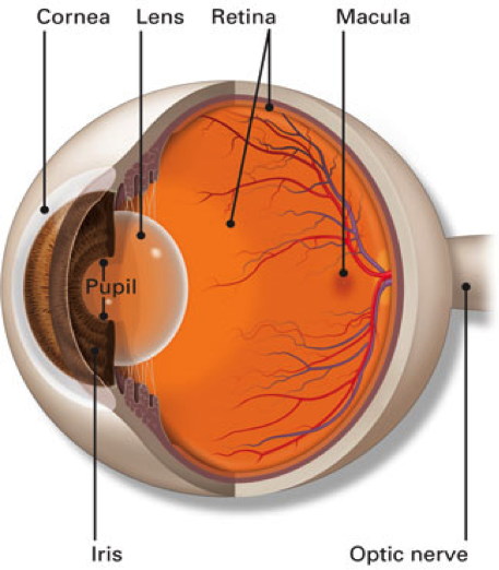
The retina is the inside lining of the back of the eye. It is composed of many layers of light sensitive nerve cells. Light entering the eye is focused on the retina and is converted to electrical impulses which are carried to the brain by the optic nerve. The centre of the retina is called the macula. It is the most sensitive part of the retina and is used for day light and colour vision. This is in contrast to the rest of the retina which is more sensitive in the dark and is mainly used for navigational black and white vision at night.

The retina is supplied by one main artery and one main vein which enter through the optic nerve and branch all over the retina to smaller arteries and veins that cross each other like railway tracks.

source – American Academy Ophthalmology

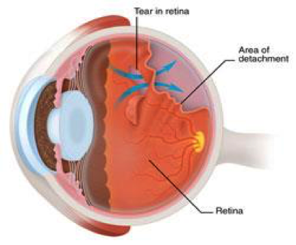
As we get older, the vitreous gel that fills the eye changes its composition and contracts causing some pulling on the retina. Most of the time it separates completely and the patient will notice some spots and lines moving across the vision (floaters), a process called posterior hyaloid detachment. In a few cases the vitreous pulling on the retina will create a retinal tear just like a cut in the wall paper. This leads to movement of liquefied gel from inside the eye cavity through the retinal tear and gradual peeling of the retina from the wall of the eye. This is what is called retinal detachment. Having lost its blood supply, the detached retina stops working which we perceive as blindness.

Retinal detachment can be either total, when the whole retina separates away from the eye wall or partial, when only a part of the retina is separated. Initially, retinal detachment starts in the periphery (macula-on detachment) causing blindness in the peripheral vision seen as a black curtain coming across from the side or from the bottom up or from the top down. If the detachment extends to involve the macula (macula–off detachment), then the patient will experience central blindness as well.

source – American Academy Ophthalmology

  • Older age
  • High myopia (short-sightedness)
  • Lattice retinal degeneration (weak areas in the retina)
  • Family history of retinal detachment
  • Some systemic conditions such as Marfan’s syndrome
  • Trauma
  • Previous retinal detachment in the other eye
  • Excessive Floaters
  • Flashes of light in the peripheral vision, more noticeable in the dark.
  • Gradual curtain like blackness progressing from the peripheral to the central vision
  • Blindness

If you experience any of those symptoms, you need to get prompt assessment.

  • Dilated retina examination: This is the standard test to check the back of your eye (retina). You will have your pupil dilated with drops so your vision will be blurry for an hour or two after the examination. Our specialist will check your eyes on the microscope (slit lamp) and use a special lens to see the details of the retina. The peripheral retina will be checked with another instrument called an indirect ophthalmoscope.
    Detailed documentation of the retinal detachment will be made including the site of the retinal tears, the status of the macula (whether it is still on or off) and whether there is evidence of stiffness of the retina in chronic cases (PVR).
  • Fundus Photography may be taken to document the extent of the retinal detachment prior to surgery.
  • OCT (optical coherence tomography); this is a fast scan of the retina centre (macula) which shows cross section details of its layers. It may be used to determine if there is fluid under the macula in cases with shallow retinal detachment.
  • Ultrasound examination: If examination of the retina is not possible because of obstructing blood in the eye cavity (vitreous haemorrhage) then an ultrasound test is used to confirm the presence of retinal detachment behind the blood.
  • Retinal Tears: If there is no retinal detachment, tears may be treated by thermal laser photocoagulation. This is usually performed at our centre while you are sitting and under local anaesthetic drops. It causes little discomfort if any, and the procedure takes only a few minutes. Laser is used in the majority of cases with weak retina (retina lattice) and tears.
    If the tears are difficult to reach with laser or large, then cryopexy is used. It is done using a freezing probe under regional anaesthetic while you are awake as a day surgery procedure.
  • Retinal Detachment: Retinal detachment usually needs prompt surgery especially if the macula is still attached (macula-on detachment). If the macula is detached (macula-off detachment) and the symptoms have been for a while, surgery can be performed over the following few days with no difference in the outcome and success rate.
    Surgery for retinal detachment has a high success rate of more than 90%. Some patients may require more than one operation. If the condition is detected and treated early, you will get the best success rate, however if the retinal detachment becomes chronic and scarring develops (PVR), then the success rate is lower.
    The goal of the surgery is to re-attach the retina to the eye wall. There are different methods used to reach this outcome depending on different criteria. These methods include:

    • Pneumatic Retinopexy: This is a relatively simple procedure usually performed at our centre and used for cases with small retinal detachment without scarring and in the top part of the retina. It includes injection of a gas bubble into the eye which will expand over the following few days. Patients will be instructed to keep their head in a certain position so as to allow the gas to seal the retinal tear thus allowing spontaneous absorption of fluid under the retina causing it to flatten. This is followed by applying laser or cryopexy to the retinal tear so as to achieve permanent scarring around it and prevent recurrent retinal detachment.
    • Scleral Buckle: This is an external surgery, the fluid under the retina is drained externally and cryopexy is used to induce scarring around the tears. A silicone band is inserted behind the eye muscles at the site of the retinal tears to achieve indentation of the eye wall and approximate the retinal tears to it thus flattening the retina. A small air or gas bubble may be used at the conclusion of surgery.
    • Vitrectomy surgery: This is an internal surgery. After excising the vitreous gel using a special cutter, the retina is flattened by a variety of methods and laser or cryopexy is applied to the tears. The eye is filled with a large gas bubble or with silicone oil at the conclusion of surgery.
      Note: if you receive gas inside the eye, you cannot fly or travel to high altitude until the gas has resolved

Want to schedule an appointment?

Call us at (+61) 3 9459 5133 or fill in the appointment form...