Age Related Macular Degeneration

The retina is the inside lining of the back of the eye. It is composed of many layers of light sensitive nerve cells. Light entering the eye is focused on the retina and is converted to electrical impulses which are carried to the brain by the optic nerve. The centre of the retina is called the macula. It is the most sensitive part of the retina and is used for day light and colour vision. This is in contrast to the rest of the retina which is more sensitive in the dark and is mainly used for navigational black and white vision at night.

The retina is supplied by one main artery and one main vein which enter through the optic nerve and branch all over the retina to smaller arteries and veins that cross each other like railway tracks.

source – American Academy Ophthalmology

Macular Degeneration is a degenerative disease of the centre of the retina (light sensitive tissue lining the inside of the eye) called the “macula” that causes progressive, painless loss of central vision. It affects the ability to see fine detail, drive, read and recognise faces. Age Related Macular Degeneration is a leading cause of vision loss in Australia amongst people 50 years of age or older.

There are two types of AMD;

  • Dry (Atrophic) Macular Degeneration
  • Wet (Neovascular) Macular Degeneration

Dry AMD is the thinning and atrophy of the cells at the macula. This is the most common type of AMD and usually causes slow gradual decline of vision over months and years. It can eventually lead to loss of central vision and legal blindness but has a better prognosis than wet AMD.

Dry Age Related Macular Degeneration (AMD)

Wet AMD is a less common but more aggressive condition. It is caused by the development of new blood vessels which grow under the retina at the macula. This leads to leakage and bleeding in the retina. Vision loss from this form of macular degeneration may be faster and more noticeable than that from dry macular degeneration.

Wet Age Related Macular Degeneration (AMD)

The top 5 risk factors of developing AMD are:

  • Being over the age of 60
  • Having a family history of AMD
  • Cigarette smoking; Current smokers have a 2-3 times higher risk for developing age-related macular degeneration than people who have never smoked.
  • Obesity
  • Hypertension (High Blood Pressure)

Macular Degeneration affects the centre of your vision but the majority of patients will maintain peripheral vision. So you may not see the details of the face of someone you are looking at directly but you can see the rest of his or her body.

Symptoms of central vision loss in AMD can include:

  • Distortion; bending straight lines like power lines
  • Central scotoma; centre of vision becomes grey or black
  • Difficulty with reading
  • Faded colours
  • Need for increasingly bright light to see up close
  • Light adaptation problems such as when entering a dimly lit room from the bright outdoors
  • Size of objects may appear different for each eye

You can check your central vision by printing the above Amsler chart:

  • Wear your reading glasses and hold the chart 30 to 40 cm ( 12 to 15 inches) away from your eyes in good light.
  • Cover one eye and look directly at the centre of the grid with the uncovered eye and keep focusing on it.
  • While looking directly at the centre dot, note whether all lines of the grid are straight or if any areas are distorted, blurry or dark.
  • Repeat this procedure with the other eye.
  • If any area of the grid looks wavy, blurred or dark, or if there is a change from the previous test, you should seek advice.

This test is useful to pick up early macular disease problems before the condition becomes advanced. Treatment and intervention is always better if diagnosis is made early to avoid untreatable damage of vision.

Although we cannot prevent getting macular denegation, there are positive steps one can take to reduce the risk of getting this condition. To reduce AMD risk

1-Quit smoking or never start.

If you smoke, you are twice as likely to develop macular degeneration compared with a non-smoker.

2- Know your family history:

If you have a close relative with AMD, you have a 50 percent chance of developing the condition. Ask your family members about it and get your eyes checked regularly. If you have early AMD, you can take further steps to reduce its progression.

3- Eat healthy

Healthy diet reduces the risk of macular degeneration.

  • Eat a well balanced diet which includes dark green leafy vegetables and fresh fruit daily
  • Eat fish twice a week
  • Choose low glycaemic index carbohydrates such as whole grain bread
  • Reduce your intake of saturated fat
  • Eat a handful of nuts a week (raw, unsalted)
  • Some people may need an extra supplement of antioxidant and vitamins 

4- Exercise

If you regularly exercise and keep normal body weight, you can have a healthier lifestyle overall. In one study, scientists found that people who exercised three times a week were less likely to develop AMD than people who didn’t exercise.

5- Check your eyes regularly

We recommend you check your retina at least once a year after the age of 60. Early signs of macular degeneration can then be detected and our ophthalmologist can tailor your management plan. If macular degeneration progresses and becomes active, early intervention is the best course to halt the disease and preserve your vision.

If you notice any of the symptoms previously mentioned then it is important to seek advise. We recommend that you get checked by an eye specialist or visit an optometrist.

The Northern Eye Centre has a specific interest and expertise in treating Age Related Macular Degeneration. We consider this condition to be an urgent case that requires immediate attention. Early diagnosis and treatment is essential to get the best outcome for your vision. You will receive a full examination of the front and back (retina) of the eye. You may also undergo immediate tests to establish the diagnosis such as with Optical Coherence Tomography (OCT) and Fundus Flourescein Angiogaraphy (FFA).

OCT of Wet AMD

FFA of Wet AMD

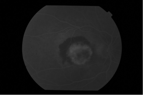
  • Treatment of Early Macular Degeneration : Macular Drusen

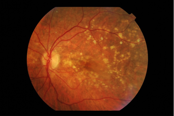
During the early stages of macular degeneration, yellow deposits called “Drusen” appear at the centre of the macula. They usually either give no visual symptoms or cause only slight difficulty with vision. There is no need for intervention at this stage.

  • Treatment of Intermediate Macular Degeneration

If the Drusen become large and plentiful, it then indicates some progression and you may be advised to take vitamin supplements.

Antioxidants and Vitamin Tablets

The Age- Related Eye Disease Study 2 (AREDS2)

This large study showed that among people at high risk for developing late-stage, or wet, macular degeneration (such as those who have large amounts of drusen or who have significant vision loss in at least one eye), taking a dietary supplement of vitamin C, vitamin E, lutein and zeaxanthin, along with zinc, lowered the risk of macular degeneration progressing to advanced stages by at least 25 percent. The supplements did not appear to provide a benefit for people with minimal macular degeneration or people without evidence of the disease during the course of the study.

The AREDS2 formula (daily dose) is: Zinc (as zinc oxide) 80 mg, Vitamin C 500 mg, Vitamin E 400 IU, Copper (as cupric oxide) 2 mg, Lutein 10 mg and Zeaxanthin 2 mg

Please note that intake of supplements does not cure macular Degeneration. It only reduces the chance of progression in some cases.


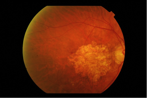
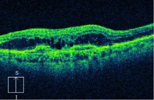
  • Treatment of Dry (Atrophic) Macular Degeneration

More common form of macular degeneration and is caused by a breakdown of cells within the macula causing thinning.

Currently there is no proven treatment for dry atrophic macular degeneration, but research is ongoing for new treatment to stop its progression. We anticipate that we will have something to offer our patients with AMD in the next few years. The intake of supplements is advised to reduce the risk of progression.

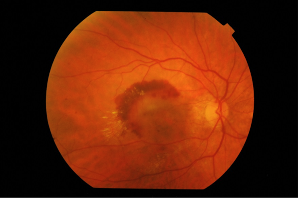
  • Treatment of Wet (Neovascular) Macular Degeneration

The most recent and common treatment of wet macular degeneration is injections with drugs called collectively anti-Vascular Endothelial Growth factors (anti-VEGF). Other modalities are also available in certain situations such as laser or photodynamic therapy.

The aim of treatment is to stop the disease and to reduce – but not to eliminate- the risk of severe vision loss. In a considerable percentage of patients vision will improve, especially if they are treated early. However it is important to know that all available treatment does not cure you from the condition.

  • Anti-VEGF injections

This is the most common and successful method of treatment of macular degeneration. We have available 3 drugs; Avastin, Lucentis and Eylea. They work in a similar fashion by blocking the effect of chemicals, called VEGF, produced in the eye which cause the new blood vessels to grow under the retina in wet macular degeneration.This will lead the leakage to dry up and stop any bleeding under the macula.

At the Northern Eye Centre, we have special expertise in treating macular degeneration and the centre is organised so that your experience is streamlined with efficiency.

Our treatment rooms are equipped so that you receive the injection whilst you are still comfortably seated in a special chair which is adjusted into position with the least inconvenience to you. The injection is performed under local anaesthesia and only takes minutes to perform.

Anti-VEGF injections need to be repeated every month initially but gradually at longer intervals. If treatment is stopped, the condition becomes active again with loss of initial benefit. It is therefore important that you follow the treatment protocol as advised by our specialist to maintain success and keep your vision sable.

o   Laser treatment

In some situations laser treatment may be necessary. Laser is a high energy focused beam of light which causes a small spot of coagulation to the retinal tissue, rather like a localised burn. This will lead to a scarring of the active new blood vessels (neovascular membrane) and stop it from further leakage. Laser is only suitable if the location of this membrane is outside the centre of the macula.

Laser treatment is available at the Northern Eye Centre. It is applied whilst you are sitting in the treatment room, it is painless and takes only a few minutes.

o   Photodynamic therapy (PDT)

This treatment involves the combination of cold low- power laser and a light activated drug called photosensitiser. The aim of the laser is to destroy abnormal ‘leaky’ blood vessels.

The procedure is performed at the Northern Eye Centre. It involves injecting the photosensitiser drug slowly into a vein in your arm. The drug will circulate in your body and reaches your retina. The low powered laser beam will then be applied to the location of the abnormal blood vessels under the macula through a special lens. The procedure is completely painless but takes some time to perform.

You will be advised to avoid strong sunlight over the following 3 days. The procedure can be repeated after a few months. PDT is currently only used in a few selected cases and can be combined with anti-VEGF injections.

This is an important question. As mentioned before, dry atrophic AMD is currently untreatable. If progressed you may become legally blind. That means your central vision has become reduced and you cannot read the top 6/60 line in the vision chart. It is only a legal term but you will still have peripheral vision. So you will still be able to walk around familiar environments such as your home but you will have difficulty seeing peoples faces or reading if the condition affects both eyes.

If you have advanced wet AMD or the injections or other treatments failed to arrest the disease progression, you may become legally blind.

In many patients, they gradually tell us they can “see again”. That is by teaching themselves to scan and use eccentric vision so they look slightly sideways if they want to look at something.

There are many organisations that we can refer you to, to help with special visual aids such as magnifiers, or even iPads. They may also provide you with home help.

  • The Bionic Eye

The Argus II retinal prosthesis, commonly known as the “Bionic Eye,” is now being tested in people with late-stage dry AMD. Until now, the device had only been used in people with retinitis pigmentosa which is a different genetic retinal disease.

The Argus II was designed to restore some useful vision in patients with significant vision loss, allowing them to see patterns of light that the brain learns to interpret as an image.

The system uses a miniature video camera stored in the patient’s glasses to send visual information to a small computerized video processing unit, which is usually worn in a pocket. This computer turns the image to electronic signals that are sent wirelessly to an electronic device implanted on the retina, the layer of light-sensing cells lining the back of the eye.

source – American Society of Retina Specialists

  • Stem cell therapy

There are clinical trials currently conducted in the UK and the US for the use of embryonic stem cell in both dry and wet age elated macular degeneration. The operation involves inserting a specially engineered patch of stem cells behind the retina to replace the diseased cells. It is hoped that the healthy stem cells will be able to sense light, restoring the central vision lost due to AMD.

It is important to understand that stem cell therapy is only under investigation at the present time and it is not approved for clinical treatment to our patients.

Want to schedule an appointment?

Call us at (+61) 3 9459 5133 or fill in the appointment form...